publications
2025
- MedIA
 Graph-based Prototype Inverse-Projection for Identifying Cortical Sulcal Pattern Abnormalities in Congenital Heart DiseaseHyeokjin Kwon, Seoungyeon Son, Sarah U Morton, and 11 more authorsMedical Image Analysis, 2025
Examining the altered arrangement and patterning of sulcal folds offers insights into the mechanisms of neurodevelopmental differences in psychiatric and neurological disorders. Previous sulcal pattern analysis used spectral graph matching of sulcal pit-based graph structures to assess deviations from normative sulcal patterns. However, challenges exist, including the absence of a standard criterion for defining a typical reference set, time-consuming cost of graph matching, user-defined feature weight sets, and assumptions about uniform node distribution. We developed a deep learning-based sulcal pattern analysis to address these challenges by adapting prototype-based graph neural networks to sulcal pattern graphs. Additionally, we proposed a prototype inverse-projection for better interpretability. Unlike other prototype-based models, our approach inversely projects prototypes onto individual node representations to calculate the inverse-projection weights, enabling efficient visualization of prototypes and focusing the model on selective regions. We evaluated our method through a classification task between healthy controls (n = 174, age = 15.4 ± 1.9 [mean ± standard deviation, years]) and patients with congenital heart disease (n = 345, age = 15.8 ± 4.7) from four cohort studies and a public dataset. Our approach demonstrated superior classification performance compared to other state-of-the-art models, supported by extensive ablative studies. Furthermore, we visualized and examined the learned prototypes to enhance understanding. We believe our method has the potential to be a sensitive and understandable tool for sulcal pattern analysis.
@article{kwon2025graph,
  title = {Graph-based Prototype Inverse-Projection for Identifying Cortical Sulcal Pattern Abnormalities in Congenital Heart Disease},
  author = {Kwon, Hyeokjin and Son, Seoungyeon and Morton, Sarah U and Wypij, David and Cleveland, John and Rollins, Caitlin K and Huang, Hao and Goldmuntz, Elizabeth and Panigrahy, Ashok and Thomas, Nina H and ... and Grant, P. Ellen and Lee, Jong-Min and Im, Kiho},
  journal = {Medical Image Analysis},
  pages = {103538},
  year = {2025},
  publisher = {Elsevier},
}

2024
- Front. Neurosci.
 The role of cortical structural variance in deep learning-based prediction of fetal brain ageHyeokjin Kwon*, Sungmin You*, Hyuk Jin Yun, and 8 more authorsFrontiers in Neuroscience, 2024
Deep-learning-based brain age estimation using magnetic resonance imaging data has been proposed to identify abnormalities in brain development and the risk of adverse developmental outcomes in the fetal brain. Although saliency and attention activation maps have been used to understand the contribution of different brain regions in determining brain age, there has been no attempt to explain the influence of shape-related cortical structural features on the variance of predicted fetal brain age. We examined the association between the predicted brain age difference (PAD: predicted brain age–chronological age) from our convolution neural networks-based model and global and regional cortical structural measures, such as cortical volume, surface area, curvature, gyrification index, and folding depth, using regression analysis. Our results showed that global brain volume and surface area were positively correlated with PAD. Additionally, higher cortical surface curvature and folding depth led to a significant increase in PAD in specific regions, including the perisylvian areas, where dramatic agerelated changes in folding structures were observed in the late second trimester. Furthermore, PAD decreased with disorganized sulcal area patterns, suggesting that the interrelated arrangement and areal patterning of the sulcal folds also significantly affected the prediction of fetal brain age. These results allow us to better understand the variance in deep learning-based fetal brain age and provide insight into the mechanism of the fetal brain age prediction model.
@article{kwon2024role, title = {The role of cortical structural variance in deep learning-based prediction of fetal brain age}, author = {Kwon, Hyeokjin and You, Sungmin and Yun, Hyuk Jin and Jeong, Seungyoon and De Le{\'o}n Barba, Anette Paulina and Lemus Aguilar, Marisol Elizabeth and Vergara, Pablo Jaquez and Davila, Sofia Urosa and Grant, P Ellen and Lee, Jong-Min and Im, Kiho}, journal = {Frontiers in Neuroscience}, volume = {18}, pages = {1411334}, year = {2024}, publisher = {Frontiers Media SA}, } - ISWA
 Visual representation learning using graph-based higher-order heuristic distillation for cell detection in blood smear imagesHyeokjin Kwon*, Seonggyu Kim, Jihye Ha, and 2 more authorsIntelligent Systems with Applications, 2024
In many real-world scenarios, including the blood smear domain, it is difficult for detection networks to achieve good performance because image annotation is usually time consuming and expensive. To address this issue, similarity-based distillation (SD) methods, considered the soft version of contrastive learning, are applied to learn a better visual representation without requiring any supervision of the downstream task. Motivated by our theoretical analysis, we treat standard SD methods as the maximization of common 1-hop neighboring key points between two queries in an attributed graph, where nodes represent query and key data points. However, such first-order graph heuristic methods that calculate the likelihood of an unseen link between target nodes by using up to 1-hop neighborhoods are normally limited by insufficient representation power and even lack of generalization ability.
@article{kwon2024visual,
  title = {Visual representation learning using graph-based higher-order heuristic distillation for cell detection in blood smear images},
  author = {Kwon, Hyeokjin and Kim, Seonggyu and Ha, Jihye and Baek, Eun Jung and Lee, Jong-Min},
  journal = {Intelligent Systems with Applications},
  volume = {22},
  pages = {200345},
  year = {2024},
  publisher = {Elsevier},
}


2022
- Front. Neurosci.
 Sparse Hierarchical Representation Learning on Functional Brain Networks for Prediction of Autism Severity LevelsHyeokjin Kwon*, Johanna Inhyang Kim*, Seung-Yeon Son, and 4 more authorsFrontiers in Neuroscience, 2022
Machine learning algorithms have been widely applied in diagnostic tools for autism spectrum disorder (ASD), revealing an altered brain connectivity. However, little is known about whether an magnetic resonance imaging (MRI)-based brain network is related to the severity of ASD symptoms in a large-scale cohort. We propose a graph convolution neural network-based framework that can generate sparse hierarchical graph representations for functional brain connectivity. Instead of assigning initial features for each node, we utilized a feature extractor to derive node features and the extracted representations can be fed to a hierarchical graph self-attention framework to effectively represent the entire graph. By incorporating connectivity embeddings in the feature extractor, we propose adjacency embedding networks to characterize the heterogeneous representations of the brain connectivity. Our proposed model variants outperform the benchmarking model with different configurations of adjacency embedding networks and types of functional connectivity matrices. Using this approach with the best configuration (SHEN atlas for node definition, Tikhonov correlation for connectivity estimation, and identity-adjacency embedding), we were able to predict individual ASD severity levels with a meaningful accuracy: the mean absolute error (MAE) and correlation between predicted and observed ASD severity scores resulted in 0.96, and r = 0.61 (P < 0.0001), respectively. To obtain a better understanding on how to generate better representations, we investigate the relationships between the extracted feature embeddings and the graph theory-based nodal measurements using canonical correlation analysis. Finally, we visualized the model to identify the most contributive functional connections for predicting ASD severity scores.
@article{kwon2022sparse, title = {Sparse Hierarchical Representation Learning on Functional Brain Networks for Prediction of Autism Severity Levels}, author = {Kwon, Hyeokjin and Kim, Johanna Inhyang and Son, Seung-Yeon and Jang, Yong Hun and Kim, Bung-Nyun and Lee, Hyun Ju and Lee, Jong-Min}, journal = {Frontiers in Neuroscience}, volume = {16}, pages = {935431}, year = {2022}, publisher = {Frontiers Media SA}, } - Front. Oncol.
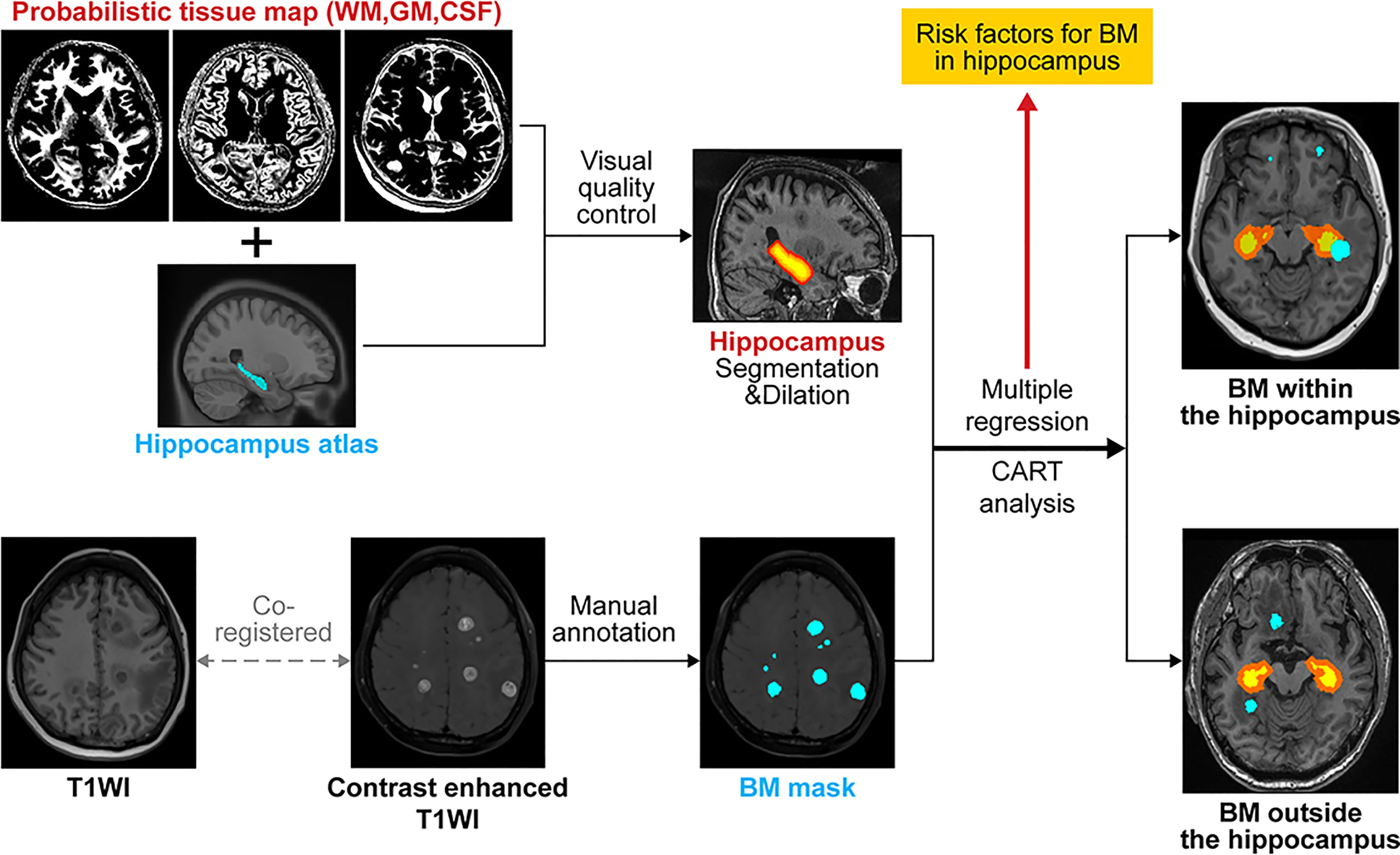 Hippocampal metastasis rate based on non-small lung cancer TNM stage and molecular markersSung Jun Ahn*, Hyeokjin Kwon*, Jun Won Kim, and 6 more authorsFrontiers in oncology, 2022
Hippocampal-avoidance whole-brain radiation therapy (HA-WBRT) is justified because of low hippocampal brain metastases (BM) rate and its prevention of cognitive decline. However, we hypothesize that the risk of developing BM in the hippocampal-avoidance region (HAR) may differ depending on the lung-cancer stage and molecular status. We retrospectively reviewed 123 patients with non-small cell lung cancer (NSCLC) at the initial diagnosis of BM. The number of BMs within the HAR (5 mm expansion) was counted. The cohort was divided into patients with and without BMs in the HAR, and their clinical variables, TNM stage, and epidermal growth factor receptor (EGFR) status were compared. The most influential variable predicting BMs in the HAR was determined using multi-variable logistic regression, classification and regression tree (CART) analyses, and gradient boosting method (GBM). The feasibility of HAR expansion was tested using generalized estimating equation marginal model. Patients with BMs in the HAR were more frequently non-smokers, and more likely to have extra-cranial metastases and EGFR mutations (p<0.05). Multi-variable analysis revealed that extra-cranial metastases were independently associated with the presence of BM in the HAR (odds ratio=8.75, p=0.04). CART analysis and GBM revealed that the existence of extra-cranial metastasis was the most influential variable predicting BM occurrence in the HAR (variable importance: 23% and relative influence: 37.38). The estmated BM incidence of patients without extra-cranial metastases in th extended HAR (7.5-mm and 10-mm expansion) did not differ significantly from that in the conventional HAR. In conclusion, NSCLC patients with extra-cranial metastases were more likely to have BMs in the HAR than those without extra-cranial metastases.
@article{ahn2022hippocampal,
  title = {Hippocampal metastasis rate based on non-small lung cancer TNM stage and molecular markers},
  author = {Ahn, Sung Jun and Kwon, Hyeokjin and Kim, Jun Won and Park, Goeun and Park, Mina and Joo, Bio and Suh, Sang Hyun and Chang, Yoon Soo and Lee, Jong-Min},
  journal = {Frontiers in oncology},
  volume = {12},
  pages = {781818},
  year = {2022},
  publisher = {Frontiers Media SA},
}


2021
- Eur. J. Neurol.
 Neural correlates of self-awareness of cognitive deficits in non-demented patients with Parkinson’s diseaseHan Soo Yoo*, Hyeokjin Kwon*, Seok Jong Chung, and 3 more authorsEuropean Journal of Neurology, 2021
Structural connectivity between and from the frontal lobes is closely associated with self-awareness of cognitive deficits in PD. Evaluating frontal structural connectivity from the early stages of PD will be important in assessing the actual cognitive and daily life performance of patients with PD.
@article{yoo2021neural, title = {Neural correlates of self-awareness of cognitive deficits in non-demented patients with Parkinson's disease}, author = {Yoo, Han Soo and Kwon, Hyeokjin and Chung, Seok Jong and Sohn, Young H and Lee, Jong-Min and Lee, Phil Hyu}, journal = {European Journal of Neurology}, volume = {28}, number = {12}, pages = {4022--4030}, year = {2021}, publisher = {Wiley Online Library}, } - NeuroImage Clin.
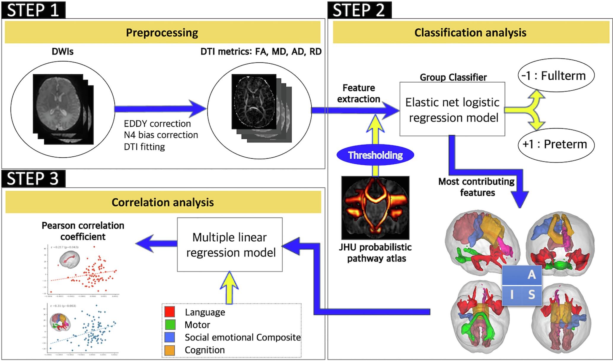 The cingulum in very preterm infants relates to language and social-emotional impairment at 2 years of term-equivalent ageHyun Ju Lee*, Hyeokjin Kwon*, Johanna Inhyang Kim, and 4 more authorsNeuroImage: Clinical, 2021
Our approach suggests that the cingulum pathways of very preterm infants differ from those of full-term infants and significantly contribute to the prediction of the subsequent development of the language and social-emotional domains. This finding could improve our understanding of how specific neural substrates influence neurodevelopment at later ages, and individual risk prediction, thus helping to inform early intervention strategies that address developmental delay.
@article{lee2021cingulum,
  title = {The cingulum in very preterm infants relates to language and social-emotional impairment at 2 years of term-equivalent age},
  author = {Lee, Hyun Ju and Kwon, Hyeokjin and Kim, Johanna Inhyang and Lee, Joo Young and Lee, Ji Young and Bang, SungKyu and Lee, Jong-Min},
  journal = {NeuroImage: Clinical},
  volume = {29},
  pages = {102528},
  year = {2021},
  publisher = {Elsevier},
}


2020
- Front. Oncol.
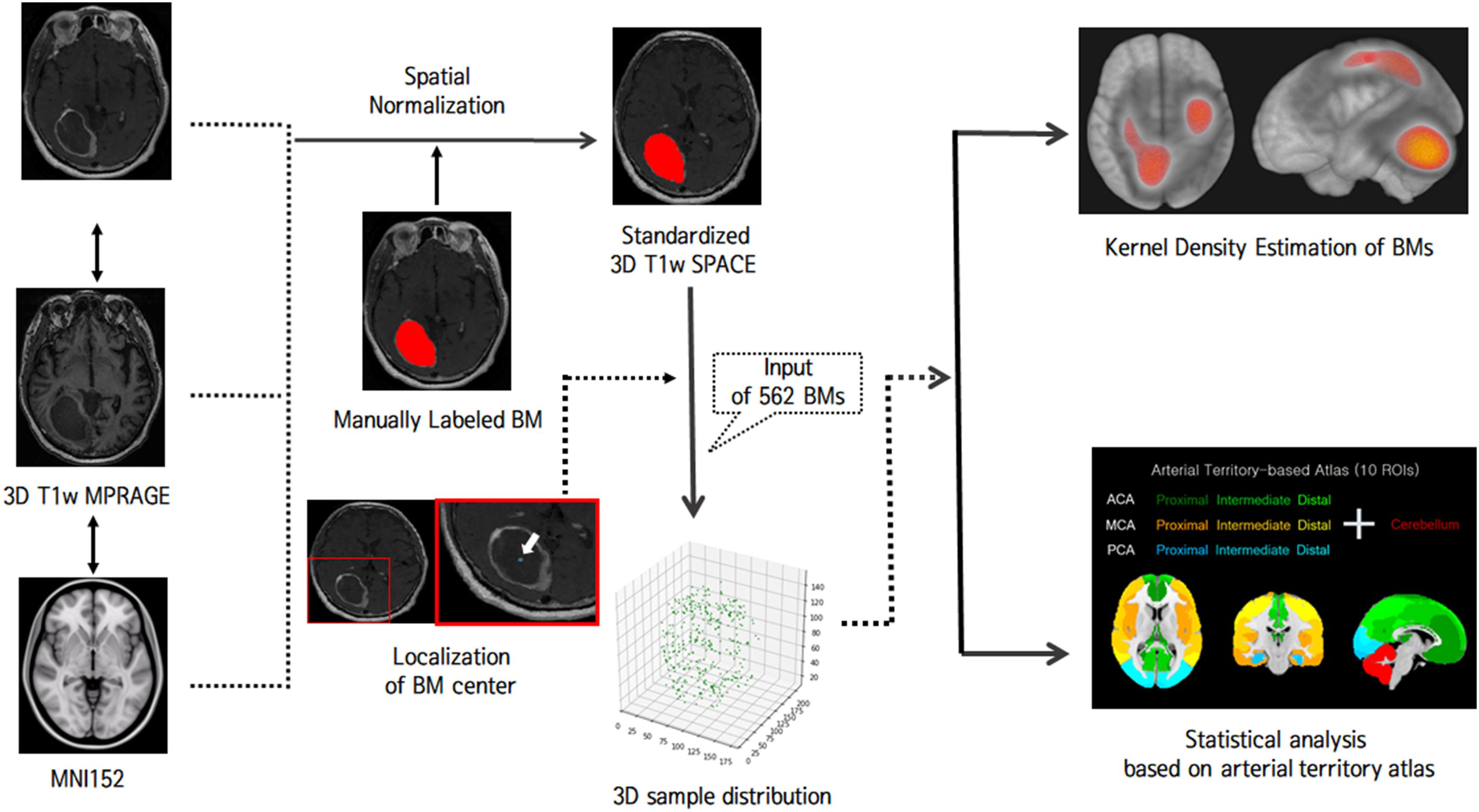 Brain metastases from lung adenocarcinoma may preferentially involve the distal middle cerebral artery territory and cerebellumHyeokjin Kwon, Jun Won Kim, Mina Park, and 6 more authorsFrontiers in oncology, 2020
Although whole-brain radiation therapy (WBRT) is the mainstay of treatment for brain metastases (BMs), the concept of saving eloquent cortical lesions has been promoted. If BMs from lung cancer are spatially biased to certain regions, this approach can be justified more. We evaluated whether BMs from lung cancer show a preference for certain brain regions and if their distribution pattern differs according to the histologic subtype of the primary lung cancer. In this retrospective study, 562 BMs in 80 patients were analyzed (107 BMs from small cell carcinoma, 432 from adenocarcinoma, and 23 from squamous cell carcinoma). Kernel density estimation was performed to investigate whether BM spatial patterns differed among lung cancer subtypes. Further, we explored more detailed subregions where BMs from adenocarcinomas occur frequently using one-way analysis of variance. Finally, we divided our cohort into those with fewer (≤10) and more (>10) BMs and evaluated whether this biased pattern was maintained across limited and extensive stages. For small cell carcinoma, BMs were biased to the cerebellum, but this did not reach statistical significance. For adenocarcinoma, BMs were found more frequently near the distal middle cerebral artery (MCA) territory and cerebellum than in other arterial territories (p < 0.01). The precentral and postcentral gyri were the most significant subregions within the distal anterior cerebral artery (ACA) and MCA territories (p < 0.01). Crus I and Lobule VI were significant regions within the cerebellum (p < 0.01). Regardless of the number of BMs, the affinity to the distal MCA territory and cerebellum was maintained. The present data confirm that BMs from lung adenocarcinoma may preferentially involve the distal MCA territory and cerebellum.
@article{kwon2020brain, title = {Brain metastases from lung adenocarcinoma may preferentially involve the distal middle cerebral artery territory and cerebellum}, author = {Kwon, Hyeokjin and Kim, Jun Won and Park, Mina and Kim, Jin Woo and Kim, Minseo and Suh, Sang Hyun and Chang, Yoon Soo and Ahn, Sung Jun and Lee, Jong-Min}, journal = {Frontiers in oncology}, volume = {10}, pages = {1664}, year = {2020}, publisher = {Frontiers Media SA}, } - Sci. Rep.
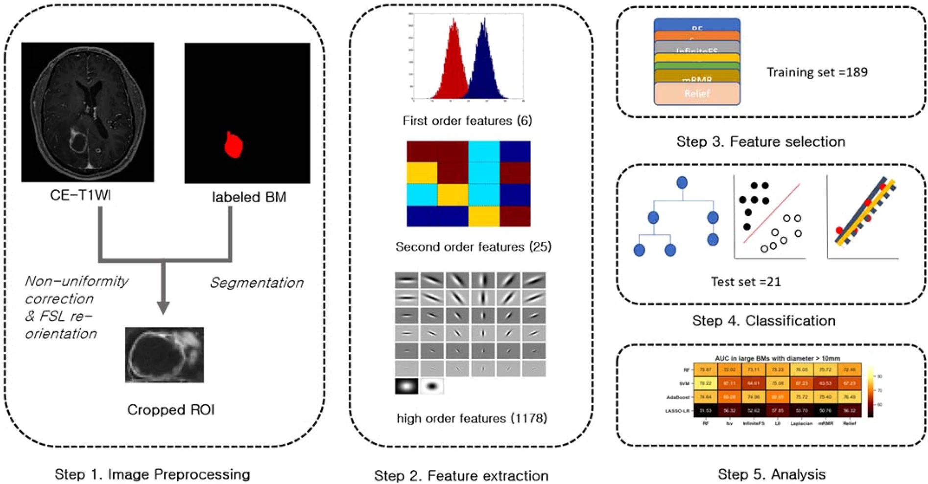 Contrast-enhanced T1-weighted image radiomics of brain metastases may predict EGFR mutation status in primary lung cancerSung Jun Ahn*, Hyeokjin Kwon*, Jin-Ju Yang, and 4 more authorsScientific reports, 2020
Identification of EGFR mutations is critical to the treatment of primary lung cancer and brain metastases (BMs). Here, we explored whether radiomic features of contrast-enhanced T1-weighted images (T1WIs) of BMs predict EGFR mutation status in primary lung cancer cases. In total, 1209 features were extracted from the contrast-enhanced T1WIs of 61 patients with 210 measurable BMs. Feature selection and classification were optimized using several machine learning algorithms. Ten-fold cross-validation was applied to the T1WI BM dataset (189 BMs for training and 21 BMs for the test set). Area under receiver operating characteristic curves (AUC), accuracy, sensitivity, and specificity were calculated. Subgroup analyses were also performed according to metastasis size. For all measurable BMs, random forest (RF) classification with RF selection demonstrated the highest diagnostic performance for identifying EGFR mutation (AUC: 86.81). Support vector machine and AdaBoost were comparable to RF classification. Subgroup analyses revealed that small BMs had the highest AUC (89.09). The diagnostic performance for large BMs was lower than that for small BMs (the highest AUC: 78.22). Contrast-enhanced T1-weighted image radiomics of brain metastases predicted the EGFR mutation status of lung cancer BMs with good diagnostic performance. However, further study is necessary to apply this algorithm more widely and to larger BMs.
@article{ahn2020contrast,
  title = {Contrast-enhanced T1-weighted image radiomics of brain metastases may predict EGFR mutation status in primary lung cancer},
  author = {Ahn, Sung Jun and Kwon, Hyeokjin and Yang, Jin-Ju and Park, Mina and Cha, Yoon Jin and Suh, Sang Hyun and Lee, Jong-Min},
  journal = {Scientific reports},
  volume = {10},
  number = {1},
  pages = {8905},
  year = {2020},
  publisher = {Nature Publishing Group UK London},
}

